Thursday, February 27, 2014

Interesting Fact About Ultrasound and Thyroid Cancer

When you hear the word cancer you think of the worst possible things. Cancer can be extremely complex, hard to treat and just about impossible to shake. Depending on the type of cancer there is a concern that the cancer may be a serious problem. Thyroid cancer is one of dangerous types of cancers that represent a major problem for patients. Thyroid cancer, like almost every other cancer, is a killer.

As with most forms of cancer diagnostic imaging plays a part in ensuring that there’s three key things:

  1. An early diagnosis.
  2. A treatment program.
  3. A response to the treatment program.

Even with diagnostic imaging processes in play there is a danger associated centered around exposure to radiation. So how does imaging help in terms of thyroid cancer? Ultrasound is proving to aid in identifying cancer in low risk patients. While the words low and risk seem to create that feel of “it can happen to anyone,” it’s fair to assume that it means something positive.

It was published in diagnosticimaging.com that “Researchers from the University of California, San Francisco, undertook a study to quantify the risk of thyroid cancer associated with thyroid nodules, based on ultrasound imaging characteristics. The retrospective, case-controlled study assessed 8,806 patients who underwent 11,618 thyroid ultrasound examinations from January 2000 through March 2005. A total of 105 patients were subsequently diagnosed with thyroid cancer.”

These kinds of studies are the kinds that create a sense of optimism among patients. This is the type of thing that serves as a step in the right direction in the battle against cancer. There is no telling if this will extend into other arenas but it’s a start.


No patient should ever feel confident about the fact that he or she is at low risk of anything. Often times some things go unseen and they actually show up much later, when it’s entirely too late to ensure that the condition, cancer or not, can be treated properly.

In the end this is the kind of study that aids in creating a greater chance for early detection. While this study covered a significant period of time, and cases, it will still draw some heat. A number of factors will still need to be considered before it can be determined if it’s completely accurate and can in turn create another route in the fight against cancer.


If you have any questions about diagnostic imaging procedures please let us know. Our dedicated team of professionals here at Clermont Radiology looks forward to answering any questions that you may have.


Charla Hurst
General Operations Manager

Charla@ClermontRadiology.com  
352-241-6100 


References: http://www.diagnosticimaging.com/ultrasound/ultrasound-images-identify-thyroid-cancer-low-risk-group

Tuesday, February 25, 2014

How Online Education May Help Radiation Concerns

Radiation is like and old friend that we want to like but can’t, at least in the world of diagnostic imaging. Radiation is a necessity when performing CT scans, Mammograms, and just about any other X-Ray procedure. The concern with radiation is not that it will hurt the patient. The real danger comes with repeat exposure, which can cause an adverse effect on the patient.


Patients with complex conditions often face dangers such as increased risks of cancer. For the last few years there has been a massive puss to go ahead and minimize said risks. Graduating radiation dosage has become and reducing it to where it doesn't compromise the image has been one way to go and in some cases it has proven effective. Even though there is something in play in order to ease concerns, radiation is still a concern.

So what’s new in the world of radiation control? The answer is online education, and so far it’s looking like a potential winner. According to an article in itnonline.com it’s been stated that “The American Society of Nuclear Cardiology (ASNC) has recently released an online education program for physicians and technologists involved in the practice of nuclear cardiology. This program offers continuing education credits and provides valuable quality control procedures and radiation safety information.”

While this particular blurb states that the online education program applies to nuclear cardiology, the development itself has some serious implications in the world of imaging as a whole. This is the type of advent that may well prove to be a game changer as it will give practitioners in general an idea of how to deal with radiation.

This isn't something that’s going to kill the radiation issue but it’s still something that helps. Likely there is going to be more in terms of research, work and ultimate results and in the end something good may come of it. Let’s see what the future holds in terms of the radiation situation.


If you have any questions about diagnostic imaging procedures, please feel free to give us a call. Our dedicated staff of professionals here at Clermont Radiology looks forward to answering any questions that you may have.


Charla Hurst
General Operations Manager

Charla@ClermontRadiology.com  
352-241-6100 


References:

Thursday, February 20, 2014

Get those Pilots Tested

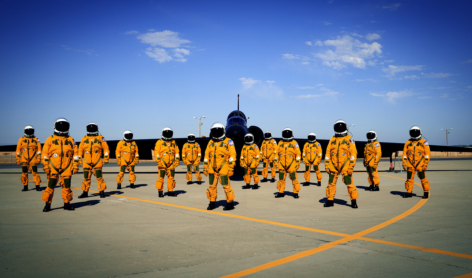
Diagnostic imaging is sometimes beyond vital in specific situations. Sometimes people in certain lines of work are put in a spot where there may be certain risks. Brain lesions are a big risk and present a big problem for patients. United States Air Force pilots are at a big risk for brain lesions. Currently the study that revealed this particular issue did not present any long term implications.


The study was done conducting MRI scans and studying existing ones as well. A brain lesion may not initially show problems but time is never really someone’s ally in this kind of situation. It’s not an easy thing to deal with especially when in the armed forces. The problem is faced more by U2 pilots. According to the article about the study “The U-2 is a high-altitude reconnaissance aircraft that maintains a cabin altitude of approximately 9000 m while operating above 21,000 m, the investigators note in their report.” It’s understandable that these pilots know the risks but these risks can hopefully be scaled back.

Risks such a decompression sickness has only gotten worst among pilots since 2006. These developments are great in the process of discovery, especially considering the fact that most conditions have some way that they can be avoided. Still, according to the article published in medscape.com : "A normal, healthy, young adult should not have very many, if any, detectable brain lesions. In the case of our U-2 pilots or divers, they are probably the result of an occupational exposure which was previously unrecognized.” The dangers of these lesions were not addressed right away but, anything in the brain can be unpredictable. As these studies develop there will hopefully be a way to keep these lesions from occurring with the development of better equipment for pilots.

If you have any questions about diagnostic imaging procedures please feel free to give us a call. Our attentive and professional staff here at Clermont Radiology looks forward to answering any questions you may have.


Charla Hurst
General Operations Manager

Charla@ClermontRadiology.com  
352-241-6100 


Tuesday, February 18, 2014

Rare Uterine Conditions, and the MRI

There are always conditions we know and conditions we do not know. Issues such as cancer, heart ailments, and kidney problems get a lot of press because it’s what people deal with on a common basis. Often times those conditions are known as complex conditions. Complex conditions are difficult to treat and sometimes the prognosis is bad depending on when they are discovered.

Uterine conditions that affect the reproductive system in women younger than their 20’s are getting better exposures through MRI scans. In an article in auntminnie.com it was stated that “MR images confirmed that women with the condition, known as Mayer-Rokitansky-Kuster-Hauser (MRKH) syndrome, may have large uteri with no conventional structure (defined as rudimentary uteri in this study) that can mimic normal postpubertal uteri. By discovering MRKH syndrome in young women, MRI can prompt the diagnosis of the disorder.”

Without a diagnosis this kind of condition can be a problem to deal with if it goes undetected for an extended period of time. Like most studies there’s always a downside and the downside is that there is a study limitation. Not every patient underwent an MRI and as a result there’s always that particular difference as far as numbers are concerned.

The article also stated that “The study also found that one or both ovaries were not in their correct location (ectopic) in 27 patients.” This can be something of a problem, especially when you consider the fact that the study was conducted using only over 60 patients. The ultimate goal of this study was to prove that MRKH Syndrome does not adversely affect the development of the uterus, even though it may affect fertility.

 For women to be able to have children, with this condition, it’s necessary to have the ovaries in the correct location, it’s actually vital. While this is one study, hopefully there will be other developments that will continue to back the results proposed by the study.

If you have any questions about diagnostic imaging please feel free to give us a call. Our dedicated and attentive team of professionals here at Clermont Radiology looks forward to answering any questions that you may have.


Charla Hurst
General Operations Manager

Charla@ClermontRadiology.com  
352-241-6100 


Thursday, February 13, 2014

Women in Radiology

When we look back at the history of women’s rights we don’t think past major events such as the suffrage movement that led to the right to vote. Women eventually would follow that up with landmark achievements. So how did women fare in the world of medicine? How did women fare in Radiology? Let’s look at some interesting facts:
  • There are still more men in radiology than women.
  • It was a woman, Madame Curie, who pioneered radioactivity research.
  •  In 1981 the American Association for Women Radiologists was born


The fact that it wasn't until 1981 that this particular association was born really shows that progress was coming along slowly. Diagnostic imaging as a whole is still a male dominated field but the AAWR made some real strides in terms of leveling the playing field in this particular specialty. The fact is that the planning stages were particularly long because prior to it’s birth there was a lot of work being put in to create stronger roles for women in terms of having associations of this type. Some of the interesting facts about the 1981 meeting that gave birth to AAWR are the following:

  • That they be accepted as equal professionals.
  • That they do not want a position they are not qualified for.
  • That the RSNA aid in the education of the public to accept the woman physician.
  • That an article appear in RADIOLOGY giving endorsement to the development of a Women's Radiological Society.
  • That there was a need to obtain mailing lists of women radiologists and a data bank for geographical location and occupation of women radiologists.

These types of requests showcased the fact that women were not seen as equals in the field of medicine and not in radiology. The creation of the AAWR was the first real step in giving women their just credit and recognition as equals in medicine and the specialty of their choosing. Women’s equality in radiology still has a long way to go, but no one can question a woman’s ability to perform a CT scan, of MRI scan based on gender. As the number of women radiologists continues to rise, so too does the already rich history of the AAWR.

If you have any questions about diagnostic imaging procedures please feel free to give us a call. Our team of dedicated professionals here at Clermont Radiology looks forward to answering any questions that you may have.


Charla Hurst
General Operations Manager

Charla@ClermontRadiology.com  
352-241-6100 


References: 

Tuesday, February 11, 2014

CT Reconstruction, The Next Generation

Diagnostic imaging processes have come a long way since the invention of the X-Ray in 1895. Reading and interpreting results has gotten easier as time has elapsed. The ability to zoom, guide, and get greater detail on an image has allowed radiologists and clinicians alike the ability to not only create a better diagnosis but also a better treatment program for patients suffering from possibly complex conditions. A new development has come about and that’s the interactive reconstruction of raw data.


Raw data with regards to the CT is a topic that’s often discussed. The definition of raw data is a simple one, there is no unique definition. To different clinicians and radiology it can mean different things. While there is that huge debate over a proper definition, work continues on the interpretation end. Dr. Elliot Siegel from the University of Maryland has stated that “Reconstruction on demand could provide the opportunity for innovation in the continuing evolution of image visualization at the workstation.” This kind of statement means that while there is a lot to be done, this process could shorten the situation.


A workstation that has the ability to reconstruct data can definitely work well. Workflow is a key element to successful imaging. It was stated that “In addition to being able to zoom in on a particular section of an image to access the full sampled data in the sinogram, this would allow radiologists to deploy particular reconstruction kernels based on specific examination type or exam indication. They could also optimize their evaluation by being able to apply multiple types of processing techniques dynamically and in real-time on different areas of the same image.”


To be able to do anything in real time improves the process to an extent that may aide in the quest for early detection and subsequent treatment, leading to a better overall prognosis.


If you have any questions about diagnostic imaging procedures please feel free to give us a call. Our dedicated team of professionals here at Clermont Radiology looks forward to answering any questions that you may have.


Charla Hurst
General Operations Manager

Charla@ClermontRadiology.com  
352-241-6100 


References: 


Thursday, February 6, 2014

Promise in treatment of traumatic brain injury

A person suffering a traumatic brain injury is likely to be in a vegetative state. This type of situation is tragic and takes a heavy and horrible toll on the families. Diagnostic imaging is taking center stage in terms of what it can do for traumatic and severe brain injuries. With a severe brain injury there is little if any response as far as the body is concerned. As time goes on the chances of recovery go from microscopic to nonexistent.

The functional MRI has allowed researchers to see certain activity in non responsive patients. It was stated that “One patient who had maintained a clinical diagnosis of vegetative state over a 12-year period prior to scanning, and also subsequent to it, was able to use attention to correctly communicate answers to several binary (yes/no) questions. In this way, the patient demonstrated that he was aware of his identity and whereabouts." This is a fascinating development that shows a possibility for really helping patients with severe brain injury.

While this kind of development doesn't mean that there is a way to fix severe brain injuries. There is a lot of hope and that’s a very good thing. It was mentioned in the article on medscape.com about this development that “fMRI may be used to communicate with patients who have a total locked-in syndrome "may lead to a paradigmatic shift such that with the further development of prostheses, these unfortunate patients, as well as patients with other forms of locked-in syndromes, will be able to open the door to end their isolation."

Isolation is horrid and can destroy the patient, and it can also destroy the patient’s family. The fact that diagnostic imaging can be such a big part of a future recovery process is something amazing. The big implication here is that the research and future technologies are going to play a major role in furthering this type of study.

If you have any questions about diagnostic imaging procedures please feel free to give us a call. Our attentive staff here at Clermont Radiology looks forward to answering all your questions and aiding you in all your diagnostic imaging needs.


Charla Hurst
General Operations Manager

Charla@ClermontRadiology.com  
352-241-6100 


Tuesday, February 4, 2014

The Brain Tumor Nightmare

Brain tumors are very difficult to deal with. Diagnostic imaging can help spot a brain tumor through early detection and aggressive and methodical treatment. Even with methodical treatment and early detection, tumors are unbelievably unpredictable. A patient can beat a tumor and face it again many years later down the line, just like the patient can live many years while still dealing with the tumor. A tumor is like an unwanted dinner guest that you have to deal in some way or another.


When dealing with brain tumors there are a few options, none of which are necessarily good. There are invasive biopsy procedures, or PET scans with a great many limitations. Ultimately the issue is not necessarily in the procedure so much as it is in the amount of discovery. Tumors are tricky but they are a lot trickier when relating to the brain.


In an article published by auntminnie.com it was stated that “An advanced MR analysis method can reveal new information on blood vessel type function, enabling physicians to determine within days whether brain tumor patients are responding to antiangiogenesis therapy.” The meaning of this is that going into greater detail will help a brain tumor. Treating tumors requires attention to detail more so than other conditions.


Hopefully the ability of physicians to tell whether or not the patient is responding to brain tumor treatment will continue to increase. Even with proposed nano technology for diagnostic imaging, there is an enormous amount of road left in so far us properly treating a brain tumor and seeing if that tumor is responding to the treatment.


The study that was conducted with regards to faster response times stated that “Patients who responded to chemotherapy survived six months longer than non-responders, a significant difference for patients with an expected survival of less than two years, according to the researchers. Quickly identifying those whose tumors don't respond would allow discontinuation of the ineffective therapy and exploration of other options.” Where chemotherapy is concerned there is always a major worry of after effects but it can help a patient.


If you have any questions about diagnostic imaging procedures please feel free to give us a call. Our attentive and knowledgeable staff here at Clermont Radiology looks forward to answering any questions you may have.


Charla Hurst
General Operations Manager

Charla@ClermontRadiology.com  
352-241-6100