Thursday, December 19, 2013

A New X-Ray Blocking Cream in Town

All physicians have some sort of cream that they use for their hands, in terms of interventional procedures. Often times procedures require some manner of diagnostic imaging to serve as a guide for the physician. When it comes to just actual procedures there is a legitimate concern in terms of exposure to radiation. If an interventional radiologist has to run multiple tests per procedure there is a concern.


Radiation exposure is not just an issue for patients; in fact it’s a big issue for radiologists and their staff as well. Doses of radiation repeated over a lengthy period of time can expose someone to risks such as cancer. 

BloXR's Ultra Blox x-ray attenuating cream, as the hand cream is known, is a mix of bismuth oxide with hand lotion. The level of protection is well backed by research and the cream did get an FDA clearance.


The advantage of the cream is that it allows interventional radiologists more time and freedom to focus on their task. Sure cream may not seem like such a big deal but when dealing with patient well being and personal peace of mind it’s vital to take every little advantage that you can.


An article about it best describes it “The procedure can last two or three hours, and the operator is getting irradiated for that length of time. Yes, it's always a concern when the patient is getting irradiated, but patients hopefully get irradiated at the most once or twice in their lifetime. The problem with clinicians is they're chronically exposed for 30 to 40 years... and it becomes potentially a long-term chronic exposure problem." Exposure really is a danger that affects clinicians.


This new cream is the kind of advancement that’s not necessarily high tech but it really helps. Not everything is a high tech innovation in particular modalities.


If you have any questions about diagnostic imaging procedures please feel free to give us a call. We here at Clermont Radiology look forward to answering any questions you may have.


Charla Hurst
General Operations Manager

Charla@ClermontRadiology.com  
352-241-6100 


References: 


Tuesday, December 17, 2013

Some Radiology History

Diagnostic imaging is full of interesting little facts that we don’t often stop to think about. Let’s look at some interesting history:
  • Radiology began as a sub specialty in the first part of the 1900’s.
  • Radiographs were made onto glass plates.
  • Film was introduced in 1918 but would not be used for years to come.
  • Society of radiographers was formed in 1920.
  • The image intensifier and X-Ray television came in the 1950’s.
  • Real time ultrasound came in the late 1970’s.


During the early days of radiology the patient had to hold a film cassette. What’s incredible is that during the early days an X-Ray could take close to twenty minutes, where now it takes almost no time at all. While radiology was thriving as time progressed there were several different developments that were somewhat adverse such as:
  • Exposure to radiation. 
  • Limited processing.
  • Contrast agents were not as advanced and while they helped, they had a long way to go.


It’s incredible to think that contrast agents or “dye” has been around for well over 100 years. Contrast agents were injected in order to create a better and more effective view of the body. Prior to the use of the image intensifier the system in place was a complex type of mirror maze that was extremely difficult to handle. Today we rely on digital technology but the truth of the matter is that they have been around for almost four decades. Some of the benefits of digital technology include:
  • Less x-ray dose can often be used to achieve the same high quality picture as with film.
  • Digital x-ray images can be enhanced and manipulated with computers giving physicians the ability to get a closer look at the images.
  • Digital images can be sent via network to other workstations and computer monitors so that many people can share the information and assist in the diagnosis creating a more effective and patient centric workflow.
  • Digital images can be retrieved from an archive at any point in the future for reference, making it easy for physicians to be able to compare cases and develop some form of effective treatment.


If you have any questions about diagnostic imaging procedures please feel free to give us a call. We here at Clermont Radiology look forward to answering any questions that you may have.


Charla Hurst
General Operations Manager

Charla@ClermontRadiology.com  
352-241-6100 


References:


Thursday, December 12, 2013

Evolution of Communication

The simplest definition of communication is the exchange of information through one means or another. Verbal communication and other non- verbal forms are sometimes complex and create difficulty. In the world of diagnostic imaging it’s no different than in the regular world. In order to better help the patient it’s vital that radiologists better communicate with clinicians.

Radiologists don’t just read an X-Ray and formulate a diagnosis. Radiologists have a responsibility to communicate properly with the clinicians and also provide results in a manner that puts the patient at ease. As procedures get more complex and require more attention, so to does the evolution of communication. A radiologist cannot just jot down a few notes and take a wild stab at a possible issue. It’s important that a diagnosis be the correct one. Often times a procedure may be repeated but in order to avoid the extra expense it is vital to get that treatment program right away.

According to Dr. Mark Flyer “During the past decade, the field of radiology has also grown exponentially, and clinicians have ordered significantly more diagnostic exams across all modalities. In today's fast-paced and consumer-driven medical environment, more demands are imposed on clinicians to deliver quick and accurate care. These increasing demands have made it difficult for them to review imaging results in a timely fashion, resulting in potentially dire consequences for their patients and medicolegal implications for their organizations.”
What Dr. Flyer is basically is stating that timeliness is vital but it cannot sacrifice accuracy. Dr. Flyer refers to a critical test results management software oriented toward the improvement of communication between radiologists and clinicians. Another interesting point brought up by Dr. Flyer was “Radiologist-clinician communication failure is a major concern in today's malpractice litigation, and it has become a major focus for improvement and regulation by the ACR. The process of reaching clinicians in today's hospital environment is often time-consuming and reduces radiologists' productivity.” Malpractice litigation is a major issue and when it can be traced back to miscommunication it makes it nearly impossible for physicians in general to be able to perform properly.
As time passes these systems will continue to improve communication, timeliness and accuracy for the patient. Ultimately the goal is to make medical practice as successful as possible and the patient satisfied.
If you have any questions about diagnostic imaging procedures please feel free to give us a call. We here at Clermont Radiology look forward to answering any questions you may have.

Charla Hurst
General Operations Manager

Charla@ClermontRadiology.com  
352-241-6100 


References:

Tuesday, December 10, 2013

Robots in the Battle

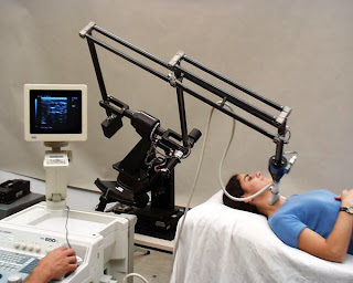
Kidney cancer is one of the worst conditions that a person can go through. Ultrasound is a commonly used tool to detect cancer in the kidneys. Through detection comes treatment and that usually can lead to the control of the condition. When it turns into something serious the results can often be very problematic and difficult. There’s a new aide in the diagnostic imaging and surgical war against kidney cancer. The new aide is none other than the robot. The Robot is not just a dance or character from Lost in Space. The robot in terms of a surgical procedure is the robotic ultrasound.

According to a related article “The kidney surgeon who performs the ultrasound robotically has direct control over the meticulous procedure, instead of having to rely on an assistant for much of the surgery.” It’s fair to assume that the Robot works as a surgeon of sorts without really performing anything outside of the ultrasound.

While there was a comparison between the use of robotic ultrasound and the use of an assistant to guide the probe, the robotic ultrasound seems to be getting a major amount of ground. The comparative study between the two methods showed similar results but there is a lot to be said for robotic ultrasound. A doctor that can fully concentrate on operating without having to worry about mistakes, at least that’s what’s being alluded to.

There is a concern that needs to be addressed and that is the concern over robotic malfunction. While this is something that was not mentioned it may be interesting to see if it’s brought up. One good thing is the fact that the ultrasound is the least invasive and harmful imaging procedure when it comes to dealing with cancer. Yes, there are many battles to be fought in the cancer front but diagnostic imaging is up to task.

If you have any questions about ultrasound or any other diagnostic imaging procedures please feel free to give us a call. We here at Clermont Radiology look forward to answering any questions you may have.


Charla Hurst
General Operations Manager

Charla@ClermontRadiology.com  
352-241-6100 


References: 


Thursday, December 5, 2013

Diagnostic Imaging Goes Small

Diagnostic imaging is its own world and it changes on a daily basis. Hybrid modalities are invented and new ways to make procedures safer are brought forward. New manners of existing modalities are released to the market and there are many attempts at going ahead and making procedures universally affordable no matter the region. So what do you know about diagnostic imaging? Here are a few things that can be told:


  • MRI stands for magnetic resonance imaging
  • CT stands for computed tomography
  • X-Ray is the gold standard and was invented over 100 years ago
  • All of the machines are fairly large

Diagnostic imaging modalities all have size and many buttons in common. When you step in to get a CT scan or you step in to get an MRI there’s something of a feeling of intimidation. Knowing that there is also some risk given the radiation that’s used, you feel a little of the nerves. So with the latest invention in diagnostic imaging there’s a great deal of surprise. The latest invention in scanning is a small molecule scanner the world’s smallest.

This particular invention is fresh and has yet to clear the massive hurdles that it must clear before being released to the market. Chances are that before this molecule scanner comes out it will be a few good years but this is amazing either way. According to the article on the invention it was stated that “Our invention allows lines to be 'written' and 'erased' much in the manner that an Etch A Sketch® toy operates. The only difference is that the smallest feature is a trillion times smaller than the children's toy, able to create conductive lines as narrow as two nanometers.” The invention will be small but the implications are gigantic.

This miniscule fix it all has what a lot of modalities lack, the ability to go molecule by molecule. The idea that the body can be read in flawless detail is huge. This could mean a different approach to cancer, heart conditions, maybe even AIDS.  Perhaps this is all too much, given this invention just came about, but it’s not bad to hope.

If you have any questions about diagnostic imaging procedures please feel free to give us a call. We here at Clermont Radiology look forward to answering any questions you may have.


Charla Hurst
General Operations Manager
Charla@ClermontRadiology.com  
352-241-6100 


Tuesday, November 26, 2013

Country Life vs City Life

Ever wonder what the difference between city life and country life is? In some cases it can be huge due in part to the following factors:

·       Nutrition
·       Quality of the environment
·       Level of exercise

Considering these factors it would seem that living in the country is far better than living elsewhere. Perhaps country living is a wonderful option but there is something about rural living that may well prove to be pretty bad for women. The fact is that a study conducted showed that woman that live in rural areas and hail from rural areas are more likely to have a lumpectomy than to be treated by way of radiation.

According to an article in healthimaginghub.com it was stated that "These study results are concerning. All women should receive guideline recommended cancer care, regardless of where they live.” This is really a statement to the fact that in some areas not all guidelines are followed as far as treating certain complex conditions. After a diagnostic imaging procedure shows there is some complex condition, there is a set of rules to follow.

Yes, rules can be bent and in the proper interest broken, but not if they harm the patient. With drivers against breast cancer being what they are, there is no reason why this statistic should exist. It’s clear that there are certain areas to explore in terms of breast cancer. While a lumpectomy avoids immediate mastectomy, there is no guarantee that said procedure will avoid need for further surgery.

Hopefully there will be futures studies conducted that reverse this discovery. Breast cancer is still a leading killer of women throughout the world, the more that’s know and explored on how to combat it properly, the better off everyone is.

If you have any questions about diagnostic imaging procedures please feel free to give us a call. We here at Clermont Radiology look forward to answering any questions you may have.


Charla Hurst
General Operations Manager

Charla@ClermontRadiology.com  
352-241-6100 


References:


Thursday, November 21, 2013

Staying Ahead of the Bump in the Head

The thing about having kids is that they always run a sort of risk. Some kids love to explore and as a result they stick the butter knife in the light receptacle. Kids travel to the ER not because they want to, just happens that they end up that way. Ultimately children heal up but sometimes they have some side effects waiting around the corner. Often times parents feel terrified when their little ones have to submit to some sort of diagnostic imaging procedure.

Seeing a minor in a big CT scanner or MRI scanner is scary. What’s scarier is the fear of things that can happen because of the high levels of radiation. While radiation is being controlled for some things it can still be too much. So is there some way that we can keep the little ones from the dangers and perils?

If little Suzie or Johnny decide to fly from a tree house there is a resolution to the fear of the scan. Little Suzie or Johnny has a bump and chances are it’s nothing more than minimal force trauma. You are afraid as a parent because you see blood coupled with a bump and you decide that you want the little one to be at their best. You go to the ER and you have the little one monitored.

A longer stay for monitoring in the ER reduces the chances of needing a scan. According to an article in healthimaginghub.com: "Every hour of observation time in the emergency department was associated with a decrease in CT rates for children whether at low, intermediate or high risk of traumatic brain injury. Furthermore, observation prior to CT decision-making for children with minor blunt head trauma was associated with reduced CT use without an observed delay in the diagnosis of significant traumatic brain injury." It’s a good sign to see that kids can be spared the emotional and possibly physical side effects of a scan.

When you take a little one to get a scan you run the risk of the following:

·       A lot of screaming
·       A major difficulty getting the kid to sit still.
·       Chance that the little one will get claustrophobic

With a longer ER visit there is a lesser chance of any difficult stays or having to engage in a diagnostic imaging procedure.

If you have any questions about diagnostic imaging procedures please feel free to give us a call. We here at Clermont Radiology look forward to answering any questions you may have.


Charla Hurst
General Operations Manager

Charla@ClermontRadiology.com  
352-241-6100 


References:

Thursday, November 14, 2013

A Faster Way to Diagnose Disease

Complex conditions are reasons for concern and worry among patients. Cancer and many other conditions require imaging for proper diagnosis and treatment. When a diagnosis takes too long to be properly established, it usually means a nearly impossible route to a cure. Diagnostic imaging is called upon to try and help avoiding this issue. MRI scans as well CT scans can really help a person through diagnosis and treatment.


Right now there is a new technology called magnetic resonance fingerprinting that may lead to faster diagnosis. This MRI technology, according to the news, “could lead to the use of full-body MRI scans that could quickly and easily detect heart disease, certain cancers and other diseases before they become difficult to treat.” It’s basically a way to not only create early detection but also detection timely enough that will allow a proper manner of treatment to be explored and used.


A huge and very optimistic outlook has been presented as it was stated, “The technology has the potential of being incorporated into annual physical examinations, with the idea that a patient could undergo a full body scan in minutes that will be able to generate a large amount of diagnostic data that can be easily interpreted.” This is the kind of technology that’s needed in order to help a patient in even greater ways.


This technology along with a quality commitment to helping eradicate disease is something big. The MRI is making huge leaps ahead that not only put cancer in the cross-hairs of the medical community but they also take greater steps against other conditions as well. The magnetic resonance fingerprinting is based on the principle, as state in the article about it, that it “works on the idea that different types of body tissue, as well as different diseases, have unique fingerprints that could help quickly diagnose specific problems.”


A patient suffering from a highly complex form of cancer may have a higher survival rate with this kind of technology in use. Perhaps a patient suffering from a condition that may have gone undiagnosed for years may discover it in time to avoid an early grave. Hopefully this technology will be successfully implemented everywhere.


If you have any questions about MRI scans or any other diagnostic imaging procedures please feel free to give us a call. We here at Clermont Radiology look forward to answering any questions you may have.


Charla Hurst General
Operations Manager
Charla@ClermontRadiology.com  
352-241-6100 


Tuesday, November 12, 2013

Watch Your Back!

You ever get a pain in your back? When you get that pain do you go to the doctor? Over a long period there seems to have been an increase in unnecessary diagnostic imaging procedures over the last several years. It’s a sad thing to see but it’s the truth. There was an article published in diagnosticimaging.com which stated that “Using information obtained from the National Ambulatory Medical Care Survey and the National Hospital Ambulatory Medical Care Survey, researchers from Beth Israel Deaconess Medical Center and Harvard Medical School in Boston studied 23,918 outpatient visits for spine problems, representing an estimated 440 million visits total over the 11-year period.” The percentages showed that testing such as MRI and CT scans improved for back pain.


The period covered 1999 to 2010 and it shows that the treatment is not only costly but misused in some occasions. When you think that the nearly 24,000 visits used were only a fraction of 440 million and what they represented as far as patterns, it’s mind-boggling. If all 440 million visits were studied it’s fair to say that the ultimate result would be a major misuse of scans for issues that are non-complex and have a fairly easy resolution.


When it comes to situations like muscle spasms, broken bones, and certain pains, not everyone agrees with using scans to determine a course of action. Dr. James Andrews doesn’t agree with using an MRI in order to determine course of action for sports related injuries. Fractures and injuries can be serious but while a bone may never be the same, an injury won’t return on its own, unlike cancer or another complex condition.


It’s vital that diagnostic imaging not be abused and misused when dealing with the patients. CT Scanners and MRI scanners along with the X-Ray all serve the purpose of helping the patient, not bleeding the patient dry. Hopefully the use of imaging unnecessarily will cease with this type of discovery. It’s unfortunate to see this type of occurrence.


As time passes and there’s an improvement on technology chances are that this practice will all but disappear entirely.


If you have any questions about diagnostic imaging procedures please feel free to give us a call. We at Clermont Radiology look forward to answering any questions you may have.


Charla Hurst General
Operations Manager

Charla@ClermontRadiology.com  
352-241-6100 


 References:


Thursday, November 7, 2013

Why a Properly Timed Scan Truly Helps

Often times we overlook the usefulness of a CT scan, and MRI, or even the occasional X-Ray. The truth of the matter is that patients don’t really like to think about these things because they are not eager to find out that they may have something worse than a common cold, a headache, or a possibly fractured limb. There is that fear of the complex condition, a cancer, something worse than that, something that will kill us if we don’t treat it.

As we age there is also the worry that a scan will likely reveal something we are not going to be able to deal with on our own, something such as Alzheimer’s disease, or epilepsy. These types of conditions are very difficult for patients to deal with on their own. With epilepsy, being completely cured is never a certainty, and Alzheimer’s disease is a condition with no cure and progressively difficult stages of deterioration.

A new study shows a possible correlation between epilepsy and the early stages of Alzheimer’s disease. While it’s not yet a certainty, this development brings about the thought that scanning for early detection may be a good concept to consider, given the serious nature of Alzheimer’s disease. According to what was published about the study itself “epileptic activity associated with Alzheimer's disease deserves increased attention because it has a harmful effect, can easily go unrecognized and untreated, and may reflect pathogenic processes that also contribute to other aspects of the illness.”

These are the kind of things that merit a concentrated effort in order to better understand what this can mean to patients that suffer or may have suffer from some form of epilepsy or another. It’s still really difficult, and in some opinions, nearly impossible to tell what causes epilepsy or why. It will be interesting to see what the correlation is between the very difficult conditions.

The study was conducted in the following way “Dr. Vossel and colleagues studied 54 patients with both cognitive impairment and epileptiform activity, and they assessed clinical and demographic data, electroencephalography (EEG) readings, and treatment responses to anti-epileptic medications.” The way this study was conducted is indicative of an attempt to further discover links between genetics and these two conditions, while this something that has yet to be proven, it is definitely something to be considered.

If you have any questions about diagnostic imaging procedures please feel free to give us a call. We here at Clermont Radiology look forward to answering any questions that you may have.


Charla Hurst General
Operations Manager
Charla@ClermontRadiology.com  
352-241-6100 


Tuesday, November 5, 2013

How Can Imaging Get Better?

Diagnostic imaging has a set of standards that are in place for the purpose of ensuring quality. X-Rays, MRI scans, and CT scans all have to meet a standard of quality. Standards are not put in place so that patients can be charged they have a real reason for existing. Patient well being is the primary concern of all physicians and accuracy in a diagnosis and treatment program are vital. When standards are not followed or are not high enough, the patient suffers. When a patient suffers there is a chain reaction that creates adverse effects for everyone involved.

It’s fair to ask, how can standards improve? Standards in imaging are handled by separate groups that try to look at issues from the patient’s perspective. Through numerous studies there is a course of action that’s suggested and subsequently implemented. In a recent article about imaging standards being expanded Wayne Forrest wrote “A number of revisions have been suggested. For example, facilities that provide CT, PET, or nuclear medicine services would be required to monitor radiation exposure levels for all staff and licensed independent practitioners who routinely work with those modalities. The commission noted that the precautions are typically addressed with exposure meters, such as personal dosimetry badges.” This is the kind of standard that applies to both patient and practitioner.

Radiation is diagnostic imaging is a concern, considering that repeated exposure can put a person at risk. Setting a standard that monitors radiation and falls in line with the new controlled dosages that rely on size, age, and condition. While this particular standard speaks more to the fact that there is a set of regulations in place for radiation, it’s a clear example of the desire to ultimately improve patient care.

Standards are not just being expanded to make sure that staff is safe. There is a push to ensure that equipment functions are specified. Uniformity and position accuracy are some of the things subject to closer scrutiny. As long as the equipment is working properly, and all regulations are being followed, the patient wins. While commissions are put together to ensure these particular standards, it’s vital for the physician to be able to tell his or her staff what’s expected in terms of quality.

If you have any questions about diagnostic imaging procedures please feel free to give us a call. We here at Clermont Radiology look forward to answering any questions that you may have. We have a great commitment to providing the best quality and highest standards in imaging.


Charla Hurst General
Operations Manager
Charla@ClermontRadiology.com  
352-241-6100 


Thursday, October 31, 2013

Recommendations for Patients at High Risk

It doesn't matter what the condition is, there are patients that are at high risk and patients at a lower risk. With cancer, breast cancer to be specific, the situation is the same. Some women are at a far higher risk than others. The level of risk may be attributed to a number of reasons from the person’s health history to environmental factors. Truthfully a single reason is hard, if not impossible to detect. When a woman is diagnosed with breast cancer, it’s vital to ensure the proper treatment avenues are followed depending on the stage of the cancer.

So if you are at a high risk for breast cancer, what do you do? Take the proper drugs for reducing the risk of breast cancer. The drugs suggested to have a strong effect are tamoxifen and raloxifene. These two drugs can seemingly reduce the risk of developing estrogen receptor positive breast cancer. The problem on the other side of the equation is always the extreme side effects of the drugs.

The issues with the two drugs mentioned include the following side effects:

  1. Blood Clots
  2. Cataracts
  3. Hot flashes

It’s important to be aware of the good and the bad but also it’s important to be aware of whether or not this is something that applies to a person. Consulting your physician to assess your risk level is a key issue. High risk of cancer does not necessarily equate high risk of side effects and vice versa. Often times these drugs may not really be an issue. If a patient is already dealing with breast cancer, diagnostic imaging procedures as well as specific treatment programs become the main players.

As far as the risk, according to an article on foxnews.com “A woman's risk for breast cancer can be assessed with an online calculator developed by the National Cancer Institute that provides an estimate based on factors such as a women's age, number of close relatives with breast cancer, and smoking and exercise habits.” Multiple issues may play a part in determining what the actual risk is.

If you have any questions about diagnostic imaging procedures please feel free to give us a call. Our team of dedicated professionals here at Clermont Radiology looks forward to answering any questions you may have and aiding you in your diagnostic imaging needs.


Charla Hurst
General Operations Manager

Charla@ClermontRadiology.com  
352-241-6100 


References:

Tuesday, October 29, 2013

Lumps: Facts and Lies

When you feel a lump in your breasts you should not allow fear to wash over you. The fact is that there are many myths and facts about lumps and it’s important to know what’s what. If you have a lump in your breasts there’s an 80% chance that it isn't cancer. There are lumps that are just cysts and are harmless. The following is a list of myths about lumps:

  • All lumps are cancer
  • If you have a mammogram that’s normal you are all set
  • Lumps are painless
  • Young girls can’t get lumps
  • Lumps are harmless if you don’t have a breast cancer history

A lump is not easy to deal with if you don’t know what the facts are. As a patient it’s vital to stay vigilant. The following list is a list of facts about lumps:

  • If a lump is detected, you need to get it looked at
  • A mammogram is a good idea
  • A biopsy on a lump may be necessary
  • If a lump is seen as benign, it’s important to make sure that you monitor it consistently
  • Possible tumors may still loom even if it’s not in the lump

It’s important for a patient to understand that keeping an eye on lumps through diagnostic imaging procedures is a smart thing to do. Anything that can develop into a complicated and complex condition is always worth investigating. Patients at risk for breast cancer also need to understand that factors such as diet, family history, and previous successful bouts with breast cancer, are not really relevant when it comes to dealing with the disease.

If you have any questions about mammograms or any other diagnostic imaging procedures please feel free to give us a call. Our team of dedicated professionals here at Clermont Radiology looks forward to answering your questions and aiding you in your diagnostic imaging needs.


Charla Hurst General
Operations Manager

Charla@ClermontRadiology.com  
352-241-6100 


References:


Thursday, October 24, 2013

Considerations For After Augmentation

Breast augmentation is not an uncommon cosmetic procedure that women tend to undergo. The purpose is to increase breast size and boost morale, at least in most cases. With breast cancer being such a big health issue there is the consideration of how it may affect women with breast augmentations. Some questions to consider center squarely on the dangers of an implant leaking, reactions to the solutions used and so on.

A vital question to ask is: Do women that have undergone augmentation need to undergo mammograms routinely? The answer to that question is a simple and emphatic yes. There is a misconception that a mammogram will not be effective or that it will not be safe for a patient. The mammogram is the gold standard of testing for breast cancer. To tell a patient to get a CT scan, MRI, or regular X-Ray, is to give the patient options outside of what’s best. The mammogram is what can best detect any cells or suspicious lesions that may lead to cancer. There are issues with a mammogram for patients with implants that center around the implant itself.

The implant may obscure the tumor and it may in turn require the patient to undergo more mammograms. As a way to keep the mammogram in mind it’s important to think about the placement of the implant. The fact is that breast cancer does not discriminate and it’s as bad for women with implants. In the case of implants there are other recommendations.

A patient may want to undergo MRI scans in order to monitor the implant itself. A rupture may make spotting a tumor more difficult and presents even more problems for the patient. Often times the MRI may actually detect the tumor. Women with implants don’t have higher incidences of breast cancer, but there chances for it are not any less either.

The important thing patients need to remember is to stay on top of monitoring themselves. If there are lumps or suspect formations, mammogram is the next logical step. Early detection is the patient’s best protection.

If you have any questions about diagnostic imaging procedures please feel free to give us a call. Our team of dedicated professionals here at Clermont Radiology looks forward to answering your questions and aiding in your diagnostic imaging needs.


Charla Hurst General
Operations Manager

Charla@ClermontRadiology.com  
352-241-6100 


References: